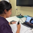
Suspicious tissue growths found during a colonoscopy may soon be able to be analysed and diagnosed in real time – avoiding the time delay of sending specimens to the lab and improving patient care.
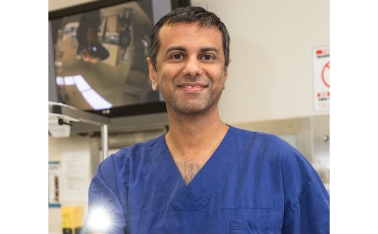
That is the hope for Professor Raj Singh, Director of Gastroenterology at Lyell McEwin Hospital (LMH).
With a grant from The Hospital Research Foundation to kickstart the project, Prof Singh and his team are developing a Computer Aided Diagnostic (CAD) system to provide a detailed analysis of tissue growths – called polyps – found during colonoscopies.
Colonoscopies are a vital step in diagnosing bowel cancer, which is 90 per cent treatable if detected early.
Any polyps found during a colonoscopy are sent to a lab for pathological analysis (called histology) and assessed as being benign, premalignant or cancerous.
“At the moment, the patient has to wait for the results to be returned,” Prof Singh said.
“We hope to be able to build our CAD system using Artificial Intelligence to predict histology in real time at more than 95 per cent accuracy. This will allow us to determine the best intervention immediately, depending on what is found.
“This will be hugely beneficial for patients and reduce the costs and complications associated with the procedure, as we won’t have to send benign polyps off for testing.”
Prof Singh is working with Associate Professor Gustavo Carneiro, a leading Computer Scientist on machine learning at the University of Adelaide, to build algorithms for the system using images of polyps collected from patients at the LMH.
About 800 images have been used so far obtaining a 70 per cent accuracy rate.
“We are continuing to collect polyps to build the computer program and once we are confident we are at a 95 per cent accuracy rate, the next step will be to trial it within an Endoscopy unit,” Prof Singh said.
Prof Singh’s innovative research hopes to improve outcomes for the many patients required to have a colonoscopy each year.